Learning is something everyone does daily—mastering new skills at work, remembering song lyrics, or following directions to new places, according to the Brighter Side of News. But behind these everyday tasks lies a complex biological process known as synaptic plasticity, a phenomenon essential for your brain's ability to adapt and store new information.
Recently, neuroscientists have discovered surprising new details about how your brain decides which synapses—the tiny connections between nerve cells—get stronger or weaker as you learn. These insights challenge earlier ideas, revealing that your brain applies multiple rules simultaneously to reshape its connections during learning.
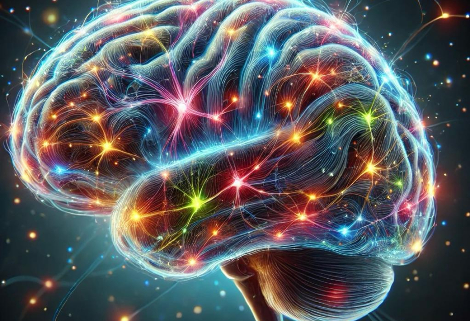
Your brain consists of billions of nerve cells, or neurons, interconnected by synapses. When you learn, specific synapses grow stronger, while others become weaker, adapting your brain's circuitry to new tasks or information.
Until recently, scientists thought that neurons used uniform rules for synaptic changes. Yet, precisely how each neuron selects the synapses for strengthening or weakening remained a scientific puzzle—known as the "credit assignment problem." Understanding this puzzle is crucial because it determines how your brain records and recalls learned information.
William “Jake” Wright, Nathan Hedrick, and Takaki Komiyama, neurobiologists at the University of California San Diego, tackled this puzzle head-on. Using advanced brain imaging methods, their groundbreaking research revealed that neurons don't follow a single rule when learning. Instead, neurons use multiple different rules simultaneously. Their findings, published in the journal Science, have profound implications for neuroscience, medicine, and even artificial intelligence.
To uncover these new insights, the researchers observed how synapses behaved during motor learning tasks in mice. Using two-photon imaging—a powerful method allowing scientists to visualize brain activity in real-time—they tracked individual synapses within layer 2/3 pyramidal neurons in the motor cortex.
These neurons have complex structures, branching into apical dendrites (extending upward) and basal dendrites (extending sideways or downward). Surprisingly, each dendrite type followed different rules for synaptic strengthening during learning.
Specifically, the team found that synapses on apical dendrites became stronger when activated alongside neighboring synapses. These synapses respond collectively to local activity patterns, strengthening connections based on what's happening nearby.
On the other hand, basal dendrite synapses strengthened when their activity coincided with action potentials—brief electrical signals traveling down neurons. In short, the apical synapses responded to neighborhood activity, while basal synapses depended on precise timing with neuron signals.
“We usually think of synaptic plasticity as uniform throughout the brain,” explained Wright, lead author of the study. “But now we see clearly that neurons use distinct rules for synaptic plasticity in different parts of their dendritic branches.”
This discovery came unexpectedly, prompting scientists to reconsider previous assumptions. Until now, research mostly indicated that synapses across neurons followed a single, uniform rule—commonly called Hebbian plasticity—where connections strengthen if both sides of a synapse activate simultaneously.
While Hebbian plasticity explains many learning processes, scientists have long suspected additional, more nuanced mechanisms exist. This study confirmed those suspicions, revealing how neurons simultaneously apply multiple plasticity rules in a compartment-specific manner.